What is Cystic Echinococcus Disease? Hydatid Cyst Formation, Symptoms, and Treatment Options
- Vet. Ebru ARIKAN

- Dec 3, 2025
- 18 min read
What is Cystic Echinococcus Disease (Hydatid Disease)?
Cystic echinococcus disease is a parasitic infection caused by the larval form of the tapeworm Echinococcus granulosus , which forms fluid-filled cysts in the internal organs. Its medical name is cystic echinococcosis or hydatid cyst disease . It is a zoonotic infection, meaning it can be transmitted from animals to humans and other animals. The disease is particularly common in rural areas and areas with a high concentration of sheepdogs and small livestock.
This parasite matures in the intestines of dogs and sheds its eggs into the environment through feces. When these eggs are ingested by grazing animals, cats, dogs, and humans, the larvae begin to form cysts in the body. The most commonly affected organs are the liver and lungs . However, spread to other organs such as the kidneys , spleen, brain, and bones is also possible.
Because hydatid cysts develop slowly, the disease can progress without symptoms for extended periods. As they grow, they put pressure on organs, disrupting their function, and can lead to serious complications in the future. When some types of cysts rupture, allergic reactions and life-threatening anaphylactic shock can occur.
Therefore, cystic echinococcus disease is a significant public health problem not only for veterinary medicine but also for human health. Early diagnosis and appropriate preventive measures are critical to reducing the risk in both animals and humans.

Echinococcus Species and Life Cycle
The primary species that causes cystic echinococcus is Echinococcus granulosus . However, other species can also cause the disease in different regions. The most important species are:
Echinococcus granulosus → Cystic echinococus (hydatid cyst)
Echinococcus multilocularis → Alveolar echinococcus, much more severe
Echinococcus vogeli and E. oligarthrus → Less common, seen in South America
For cystic echinococcus disease to occur, the parasite must complete its life cycle. This cycle is based on the dog-sheep model, but many mammals can serve as intermediate hosts.
Stages of the Life Cycle
Adult Parasite (Dog Intestine) The adult form of the parasite lives in the small intestine of dogs. The eggs are spread into the environment through the dog's feces.
Egg Stage (External Environment) Eggs can survive for weeks in the external environment. They can attach to soil, grass, water, and animal fur.
Infection to Intermediate Hosts (Human, sheep, goats, cattle, cats, dogs, etc.) When ingested, the eggs hatch in the intestine and the larval form emerges.
Oncosphere Migration (Through Blood and Lymph) Larvae penetrate the intestinal wall, mix with the blood, and are carried to the target organs.
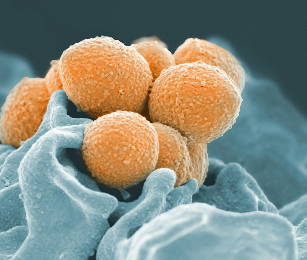
Cyst Development (Liver, Lung, Other Organs) The larva settles in the organs and forms a fluid-filled structure called a hydatid cyst. The cyst grows and produces many new larval individuals called "protoscoleces" inside it.
Infection of Dogs: If the offal of sheep, goats, cattle or other animals that serve as intermediate hosts is eaten raw by the dog, the protoscoleces in the cysts turn into adult parasites in the dog's intestine and the cycle begins again.
Clinical Significance of the Life Cycle
Cyst formation progresses very slowly and can lead to serious organ damage over the years.
The ability of eggs to survive in the environment for a long time makes transmission easier.
If left unchecked, the dog-human-animal cycle continues unabated.
Causes and Transmission Mechanism of Cystic Echinococcus Disease
Cystic echinococcus disease is caused by ingestion of Echinococcus granulosus eggs. The source of these eggs is infected dog feces . Therefore, the primary mechanism of transmission is environmental contamination.
Major Sources of Contamination
Grass and Soil Contaminated with Dog Feces: Eggs can be found attached to grass. Grazing small livestock are easily infected.
Dogs Eating Raw Offal: When raw offal such as liver and lungs from infected sheep or cattle is fed to the dog, the dog acquires the parasite and begins shedding eggs.
Eggs Sticking to Hands, Nails and Hair If hand hygiene is not provided after contact with the dog, people can ingest the eggs without realizing it.
Polluted Water Sources: Especially in rural areas, puddles and irrigation canals may carry eggs.
Pet Hair: Hair that comes into contact with dog feces may carry eggs.
Infection with humans
People usually become infected through:
Consumption of unwashed raw vegetables and fruits
Frequent contact with dogs and poor hand hygiene
Drinking dirty water
Dealing with contaminated soil in rural areas
Handling or contact with infected offal
Contagion in Cats and Dogs
Dogs: Infected when infected organs (especially liver and lungs) are eaten raw.
Cats: Direct transmission is very rare, but they can acquire eggs from the environment.
Pathophysiology of Contamination
The egg enters the gastrointestinal tract.
The larva, called an oncosphere, emerges and penetrates the intestinal wall and passes into the vascular system.
The liver is the most commonly affected organ because it is the first filtering organ; the lungs come second.
Here the larva develops into a cyst over the years.
The protoscoleces within the cyst can give rise to hundreds of new parasites.

Symptoms and Clinical Findings of Cystic Echinococcus Disease
Cystic echinococcus disease (hydatid cyst) causes clinical symptoms due to cysts formed in organs by Echinococcus granulosus larvae. However, these symptoms appear very slowly , as hydatid cysts grow over months to years, compressing organs and causing dysfunction. Therefore, the disease is often diagnosed late.
1. The intensity of symptoms depends on the location and size of the cyst.
Each organ can produce different symptoms. In the early stages, there may be no symptoms at all, and many cases are discovered incidentally during imaging.
2. General Clinical Symptoms
Weakness
Weight loss
Anorexia
Pain or feeling of pressure in the abdominal area
Respiratory distress (in lung involvement)
Cough or phlegm
Sudden hypersensitivity reactions (in case of cyst rupture)
3. Symptom Development Process
Slow-growing cysts → Silent for months to years
When organ pressure occurs → Dysfunction
When rupture (cyst burst) occurs → Shock, allergy, anaphylaxis
4. Differences in Symptoms According to Types
In ruminants, symptoms are often not noticeable from the outside; low productivity is the most important finding.
Gastrointestinal signs, abdominal pain, and elevated liver enzymes are more common in dogs .
As the cyst size increases in humans , the clinical picture becomes more evident.

Organs Where Hydatid Cysts Are Most Commonly Seen
In cystic echinococcus, the location of the cysts is the most critical factor determining the clinical presentation of the disease. Once the larvae enter the bloodstream, the cysts concentrate in the liver, the body's "first filtering organ," and then in the lungs. However, they can also spread systemically to other organs.
1. Liver (most common location with 60–70%)
Liver cysts cause symptoms through pressure and bile obstruction:
Pain in the upper right abdomen
Hepatomegaly (liver enlargement)
Jaundice
Digestive problems
Abdominal swelling depending on the size of the cyst
2. Lung (20–30%)
Lung cysts affect breathing:
Cough
Chest pain
Shortness of breath
Bloody sputum (in advanced cases)
Feeling of pressure in the chest
Pulmonary involvement may be seen more frequently in children.
3. Other Organs (Rarer)
Spleen → Upper left abdominal pain
Kidney → Side pain, difficulty urinating
Brain → Headache, neurological findings, seizures
Bone → Pain, fracture formation, local swelling
Heart → Heart rhythm disturbances (very rare)
Muscle tissue → Palpable soft swellings
4. Multiple organ involvement
In some cases, more than one organ may be affected at the same time. The presence of multiple cysts makes the clinical picture more severe.
How Does Organ Damage Develop in Cystic Echinococcus Disease?
The organ damage caused by hydatid cysts depends on the cyst's location, size, growth rate, and intracystic pressure. Echinococcus granulosus larvae do not directly destroy tissue; the primary damage is caused by the mechanical pressure of the cyst and the immune system's inflammatory response.
1. Mechanical Stress Damage
As the cyst grows:
Compresses the organ from the outside
Impairs organ function
Puts pressure on adjacent tissues
Blocks bile ducts (in liver cysts)
Decreases lung capacity
This pressure can lead to increased intra-abdominal pressure, difficulty breathing, or organ displacement.
2. Bile Duct and Vein Obstruction
In liver cysts:
Bile flow is disrupted → jaundice
Capillaries become blocked → local ischemia
Fibrosis develops on the liver surface
Portal vein pressure may increase
If bile duct obstruction becomes chronic, it may progress to liver failure.
3. Lung Damage
Lung cysts:
Loss of expansion in the lung lobes
Respiratory surface area reduction
Pneumothorax (cyst rupture)
May create a risk of secondary infection.
4. Rupture of the Cyst (Bursting)
When the cyst bursts:
Antigens in the cyst mix with the blood
Risk of anaphylactic shock arises
Cyst contents spread into the abdominal cavity → “secondary hydatidosis”
This is a situation that is both immediate and carries a fatal risk.
5. Chronic Inflammation
A connective tissue reaction develops around the cyst wall.
Fibrosis
Hardened tissue
Loss of organ function: This process can progress over years and cause permanent damage.
Clinical Course of Cystic Echinococcus Disease According to Types
The clinical course of cystic echinococcus disease varies depending on many factors, including the infected species , immune status , the amount of eggs ingested , and the cyst's location . The same parasite can produce completely different clinical presentations in different species.
1. Clinical Course in Sheep and Goats
Sheep and goats are the classical intermediate hosts for Echinococcus granulosus. Clinical signs are usually insidious and mild:
Significant weight loss
poor condition
Mild loss of appetite
Decreased liver efficiency due to fibrosis
Sudden deaths in some herds (in case of multiple cyst load)
Hydatid cysts are usually noticed during slaughter. Economic losses are high in these species.
2. Clinical Course in Cattle
Cattle are more resistant to infection. Cysts can be mostly sterile and tend to grow slower.
Long-term asymptomatic
Silent growth in the liver and lungs
Chronic productivity loss
The emergence of cysts after cutting
It is usually subclinical; advanced cases are rare.
3. Horses and Other Large Mammals
Although infection is rare in horses:
Fatigue
Performance degradation
Weakness
Mild abdominal pain
Symptoms such as may be observed. Liver involvement is more commonly observed.
4. Clinical Course in Dogs
Dogs are the main host species of the parasite. Therefore, the adult parasite lives in the dog's intestine and often does not show any symptoms .
Egg shedding through asymptomatic feces
Rarely vomiting, loss of appetite, abdominal discomfort
The risk of environmental contamination is very high in pet dogs.
The real clinical danger to dogs is not the cyst itself, but the continuation of the anesthetized life cycle.
5. Clinical Course in Cats
Cats are rarely susceptible to Echinococcus infection. Even if they do ingest eggs, they often:
Larval development does not occur
Cyst formation is extremely rare
Therefore, they are of very low clinical significance for cats, but they may play a minor role in the chain of transmission.
6. Clinical Course in Humans
Humans become intermediate hosts accidentally. The clinical picture is slow and progressive:
Stomach ache
Cough and shortness of breath
Weight loss
Jaundice
Organ dysfunction due to cyst size
The most serious risk in humans is anaphylactic shock and spreading new cyst formations as a result of cyst rupture.
Breeds Prone to Cystic Echinococcus Disease – Table Format
The table below shows the species susceptible to cystic echinococcus disease and their susceptibility levels. (By convention, the table has three columns: Breed / Description / Susceptibility Level)
Cystic Echinococcus Predisposition Table
Race / Species | Explanation | Level of Predisposition |
Sheep | Main intermediate host; heavy infection with high egg intake | A lot |
Goat | More resistant than sheep, but chronic course is common | Medium–High |
Cattle | Infection is usually silent; cysts are often sterile | Middle |
Horse | Rare; predominantly liver involvement | Low–Medium |
Dog | The main host does not cause clinical symptoms but is the center of the transmission chain. | A lot |
Cat | Very rare; low clinical significance | Little |
Person | Accidental intermediate host; risks serious organ damage | Medium–High |
This table shows that the disease has different clinical significance in both animals and humans.
Diagnosis of Cystic Echinococcus Disease (Serology, Imaging, PCR)
Diagnosis of cystic echinococcus disease is made by directly imaging the cysts formed by the parasite or by measuring the body's immune response to the parasite. Due to the slow progression of the disease, diagnosis often requires a multimodal approach.
1. Clinical Examination
Liver enlargement
Sensibility
Difficulty breathing (lung cyst)
Weight loss
General findings, including poor physical condition, are considered. However, they are not diagnostic in themselves.
2. Serological Tests (ELISA, IHA, IFAT)
Serology is one of the most frequently used methods in the diagnosis of cystic echinococcus.
Detects antibodies against the parasite
It can give positivity even in the early stages
Invaluable in screening and herd health management
However, the disadvantage of serological tests:
Unexplained positivity (past infection)
Low susceptibility in some animal species
Therefore, serology is often evaluated in conjunction with imaging.
3. Imaging Methods
Ultrasonography
It clearly shows the size, wall structure and internal structure of liver cysts.
It is the first choice method for cysts in organs other than the lungs.
X-ray
It is a valuable screening tool in lung cysts.
Radiological opacities formed by cysts may be seen.
CT / MRI
Used in human cases and situations requiring advanced imaging
If the cyst is complex, it provides the most detailed image.
4. Stool Examination
It is possible to see adult parasite eggs in the stool of dogs; however, special techniques may be required because the eggs are microscopically similar to other tapeworm eggs. (Eggs are not seen in the stool of intermediate hosts carrying cysts.)
5. PCR and Molecular Tests
Detects parasite DNA from blood, cyst fluid, or tissue samples
Provides very high sensitivity
It is used especially in research and advanced diagnostic centers.
Hydatid Cyst Treatment: Surgery, Antiparasitic Drugs, and Monitoring Protocols
Treatment for cystic echinococcus is determined by the location , size , and number of cysts, the risk of complications , and the patient's general health. The primary goal of treatment is to both halt the cyst's growth and eliminate the risk of rupture.
Treatment consists of three main components: surgery , medical (antiparasitic) , and dynamic monitoring .
1. Surgical Treatment (Gold Standard)
Surgery is preferred for cysts that are large, create pressure, carry a risk of infection, or cause complications. The most commonly used methods are:
Cystectomy: Complete removal of the cyst.
Pericystectomy: Removal of the cyst wall along with the surrounding tissue.
PAIR method (Puncture–Aspiration–Injection–Reaspiration): Emptying the cyst with a needle, filling it with scolicidal agents and emptying it again. It is especially used in the minimally invasive treatment of liver cysts.
Surgical success rate is high, but it is important to select the appropriate case.
2. Antiparasitic Drug Treatment
Drug therapy may be used in cases where surgery is not possible, where there are multiple cysts, or where cysts are small.
Albendazole (most commonly used) Stops the cyst from growing and kills the protoscoleces within. Long-term treatment is required (usually 1–3 months or longer).
Mebendazole can be used as an alternative but is not as effective as albendazole.
Drug therapy is often combined with surgery: Albendazole is administered for 1–2 months before and after surgery. This both reduces cyst viability and reduces the risk of recurrence.
3. Follow-up and Post-Treatment Control
After treatment:
Ultrasound and radiological follow-up
Liver enzyme tests
Blood values (especially due to the effect of albendazole on the liver)
Monitoring the risk of cyst regrowth
It must be done at regular intervals. In chronic cases, the treatment process may take months.
Complications and Prognosis in Cystic Echinococcus Disease
Complications of hydatid cysts vary depending on the size of the cyst and the organ it is located in. Some complications can be mild, while others can be life-threatening.
1. Cyst Rupture (Burst)
It is the most serious complication.
Spread of cyst contents into the abdominal or chest cavity
severe allergic reaction
Risk of anaphylactic shock
Beginning of new cyst formations (secondary hydatidosis)
This situation requires immediate intervention.
2. Bile Duct Obstruction (Liver Cysts)
Impaired bile flow
Jaundice
Inflammation of the bile ducts (cholangitis)
Liver dysfunction
The cyst may press on the bile ducts or its contents may leak into the bile ducts.
3. Lung Complications
Respiratory restriction
Shortness of breath
Opening of the cyst into the bronchus
Bloody sputum
Pneumothorax (opening of the cyst into the air space)
These situations can lead to progressive and serious conditions.
4. Secondary Bacterial Infections
Damage to the cyst wall or leakage of its contents may allow bacteria to settle, leading to the risk of liver abscess or systemic infection.
5. Complications in Spine and Bone Involvement
When the cyst settles in the bone:
Pathological fractures
Pain
Progressive deformation
Serious problems such as may occur.
6. Prognosis
Early diagnosis + correct treatment: Generally good
Large and multiple cysts: Medium
Rupture + anaphylaxis: Dangerous/bad
Bone or brain involvement: Causes serious prognostic problems
The most critical factors determining the prognosis in cystic echinococcus disease are the size of the cyst and the timing of treatment.
Home Management and Nutritional Recommendations for Cystic Echinococcus Disease
Although cases of cystic echinococcus in pets are relatively rare, home care is vital during the treatment process.
1. Liver-Friendly Diet
If the hydatid cyst affects the liver:
Medium protein, high-quality food
Low-fat formulas
Fiber-rich foods that facilitate digestion
A diet rich in antioxidants is recommended.
2. Immune System Supporting Supplements
With veterinary approval:
Omega-3 fatty acids
Vitamin E
Silymarin
SAMe may support liver cell regeneration.
3. Performing Exercise Gradually
Due to the pressure of the mass, the animal may tire quickly. Exercise:
Short
Controlled
Excessive exertion should be avoided.
4. Regular Veterinary Checkups
After treatment:
Ultrasound monitoring
Blood tests
Re-drug protocol if necessary
should be monitored closely.
5. Pay Attention to Hygiene Rules
Although the adult form in dogs may not show any symptoms, eggs may be shed in the feces. For this reason:
Regular collection of feces
Keeping dog grooming areas clean
Paying attention to hand hygiene
It is very important to break the chain of infection.
6. Herd Management
In sheep and goat farms:
Offal should not be given to dogs
Regular antiparasitic applications
Pasture control
Shelter cleaning protocols
should be done.
Cystic Echinococcus Disease in Cats and Dogs: Differences Between Species
Cystic echinococcus disease presents with different clinical features in cats and dogs. The source of the disease , Echinococcus granulosus, develops its adult form primarily in the intestines of dogs . Therefore, dogs play a critical role in the natural life cycle of the disease, while cats play a much lesser role.
1. Clinical and Transmission Features in Dogs
The dog is the definitive host species of the parasite.
The adult form of the cyst lives in the dog's small intestine.
Most of the time, no clinical symptoms are observed .
The dog excretes large amounts of eggs in its feces.
These eggs easily contaminate the environment, soil, grass and water.
Thus the cycle of sheep-dog-human continues.
The most important problem for the dog is not the disease caused by the cyst, but rather that it is the main source of the chain of infection. Infection in domestic dogs most often develops through the feeding of raw offal.
2. Clinical and Transmission Features in Cats
Cats are much more resistant to parasites. Even if they get eggs:
Most larvae fail to develop,
Cyst formation is very rare,
The parasite cannot complete its life cycle in cats.
Cases of hydatid cysts in cats are extremely rare in the medical literature. Therefore, cats are not an "infectious species" like dogs.
3. Diagnostic Differences
In dogs, diagnosis can be made by fecal egg testing and PCR.
Since cyst formation is rare in cats, imaging methods (ultrasound, CT) are more decisive.
The sensitivity of serological tests may vary depending on the species.
4. Treatment Differences
In dogs, the aim is often not to treat the cyst but to stop egg shedding in the environment; therefore, regular antiparasitic application is very important.
In cats, treatment is reserved for the rare cases of liver cysts and more limited clinical guidance is available.
In conclusion, while the dog is at the centre of the disease, the cat is a species that is rarely infected and carries a low clinical risk.
Protection from Cystic Echinococcus Disease and Preventive Health Strategies
The most effective solution against cystic echinococcus is prevention , not treatment. Because interrupting the parasite's life cycle dramatically reduces the risk of disease in both animals and humans.
Prevention strategies are summarized under three main headings: animal health , environmental hygiene , human health .
1. Protection for Dogs
Raw offal should never be given to dogs.
Liver and lung tissues of infected animals should be destroyed.
Dogs should be given regular antiparasitic treatments (praziquantel, etc.) .
Dog feces should be collected daily to prevent contamination of the environment.
Control and regular treatment of stray animals is important for public health.
2. Protection of Farm Animals
Regular fasciolicide and tapeworm treatments should be carried out to reduce contamination of pasture.
Infected offal should not be fed to dogs during slaughter.
Hygiene protocols should be implemented on farms and fecal management should be done regularly.
Newly acquired animals should be quarantined and checked.
3. Environmental Control Measures
Eggs released into the environment can remain viable for a long time, so:
Water must be purified
Animal feces should not be left near irrigation channels.
Hygiene should be maintained in agricultural areas
Offal waste should be removed from the environment
4. Protection for Human Health
Raw watercress and plants of unknown origin should not be eaten.
Raw vegetables and fruits should be washed thoroughly.
Hand hygiene should be taken into consideration in rural areas.
Water of unknown origin should not be drunk.
Hands should be washed after contact with dogs.
5. Community-Level Prevention Programs
Regular veterinary checkups
Farm training programs
Antiparasitic campaigns by municipalities
Strengthening environmental waste management policies
When these strategies are implemented together, the incidence of hydatid cysts decreases dramatically.
FAQ - Cystic Echinococcus (Hydatid Cyst)
What is cystic echinococcus disease and how does it occur?
Cystic echinococcus is an infection characterized by fluid-filled cysts formed in the liver, lungs, and other organs by the larval form of the parasite Echinococcus granulosus . After ingestion, the parasite eggs hatch in the intestine, where the larvae enter the bloodstream and migrate to the liver or other organs, where they form cysts over time. These cysts gradually enlarge and cause serious organ damage.
Can cystic echinococcus disease be transmitted from animals to humans?
Yes. The disease is zoonotic. Eggs are spread in the environment through dog feces and can be ingested by humans through unwashed vegetables, contaminated water, or dirty hands. Therefore, the disease is of both veterinary and public health importance.
In which organs is hydatid cyst most common?
Cysts are most commonly seen in the liver (60–70%) and lungs (20–30%). Less commonly, they can also develop in the spleen, kidney, brain, bone, heart, and muscle tissue. Clinical symptoms vary depending on the organ where the cyst is located.
What are the symptoms of hydatid cyst?
Symptoms vary depending on the organ involved and the size of the cyst. Liver involvement can cause abdominal pain, nausea, jaundice, and an enlarged liver. Lung cysts can cause cough, shortness of breath, and chest pain. General symptoms include weight loss, fatigue, and loss of appetite.
Does cystic echinococcus disease show symptoms in the early stages?
Generally, no. Hydatid cysts grow slowly and may not cause symptoms for long periods. Symptoms usually appear when the cyst grows large enough to interfere with organ function.
Can dogs get cystic echinococcus?
Dogs are the primary hosts, meaning the adult form of the parasite lives in the dog's intestines. Dogs often show no symptoms but shed large numbers of eggs into the environment through their feces. The greatest danger for dogs is not the clinical disease itself, but their central role in the transmission chain.
Can cats carry cystic echinococcus?
Cats are remarkably resistant. Even if they ingest eggs, larval development and cyst formation are often absent. Clinical disease is extremely rare, so cats pose a low epidemiological risk.
How is cystic echinococcus transmitted?
The only way to contract Echinococcus is by ingesting eggs. These eggs are typically found in soil, grass, water, or vegetables contaminated with dog feces. Failure to wash hands after contact with dogs is also a significant risk factor.
Can cystic echinococcus disease be fatal?
Yes. If the cyst ruptures, the contents of the cyst may leak into the bloodstream, causing anaphylactic shock. Large cysts can also disrupt organ function, leading to serious or even fatal complications.
How is hydatid cyst diagnosed?
Diagnosis is made using serology, ultrasound, CT/MRI, X-ray, and PCR. Ultrasound is the most common diagnostic tool for liver cysts. X-ray and advanced imaging are preferred for lung cysts. Serologic tests are important for early diagnosis by detecting the presence of antibodies.
Can a stool test diagnose a hydatid cyst?
Eggs are not observed in the feces of intermediate hosts (humans, sheep, goats, cattle, and cats). Only dogs carrying the adult parasite in their intestines can have eggs in their feces. Therefore, a stool test can be used to detect infection in dogs.
Is surgery necessary for the treatment of hydatid cyst?
Surgery is the gold standard for cysts that are large, compressive, carry a risk of infection, or have a high risk of complications. Antiparasitic medication can be used for small cysts or for patients who are not suitable for surgery. The PAIR method is also an effective, minimally invasive option in suitable cases.
What medications are used in the treatment of hydatid cysts?
The most commonly used medication is albendazole . It stops the cyst from growing and kills the larval structures within. Treatment can be prolonged (usually 1–3 months or more). Mebendazole can be used as an alternative in some cases.
Does cystic echinococcus disease recur?
Yes. If the cyst is not completely removed or if it ruptures and spreads into the abdomen, it may recur. Furthermore, new infections may occur as long as environmental contamination persists. Regular follow-up after treatment is important.
What happens if a hydatid cyst ruptures?
A ruptured cyst is a serious emergency. If the cyst contents leak into the bloodstream, an allergic reaction, severe pain, intra-abdominal dissemination, and anaphylactic shock can occur. This condition can be life-threatening and requires immediate medical attention.
What is the growth rate of hydatid cyst?
Cysts usually grow slowly over months or years. However, in some cases, growth can accelerate. The rate of cyst growth varies depending on the type, age, immune status, and organ location.
What are the symptoms if hydatid cyst is seen in the lung?
Cough, chest pain, shortness of breath, difficulty expectorating sputum, and sometimes bloody sputum may occur. Large cysts can severely reduce lung capacity.
What are the symptoms if a hydatid cyst is seen in the liver?
Pain in the upper right abdomen, jaundice, indigestion, an enlarged liver, and difficulty digesting are common symptoms. Jaundice becomes more pronounced if bile duct obstruction develops.
What are the symptoms of cystic echinococcus in animals?
In ruminants, it usually progresses silently, with weight loss and decreased productivity. Adult parasites are asymptomatic in dogs. Clinical signs are very rare in cats. Cysts in the liver and lungs are often discovered incidentally during slaughter.
How do dogs spread this disease?
When dogs eat infected offal, they carry the adult parasite in their intestines and shed eggs in their feces. These eggs contaminate grass, soil, water, and other surfaces, becoming a source of infection for other animals and humans.
Can hydatid cyst be treated at home?
No. A hydatid cyst is definitely not a health problem that can be treated at home. It requires surgery, antiparasitic treatment, and professional medical monitoring. Home care is supportive only.
Can cystic echinococcus disease be diagnosed with ultrasound?
While ultrasound is highly effective in diagnosing liver cysts, it does not always provide a definitive diagnosis. The most accurate approach is to evaluate it in conjunction with CT/MRI and serological tests.
What should be done to prevent hydatid cyst?
Regular antiparasitic treatment of dogs, avoiding raw offal, cleaning the environment, washing fruits and vegetables, consuming safe water, and maintaining good hand hygiene are the cornerstones of prevention. These strategies break the chain of transmission.
How do I know if I have cystic echinococcus disease?
Symptoms often appear late. Abdominal pain, cough, weight loss, elevated liver enzymes, or incidentally detected cysts during imaging suggest infection. Imaging and serology are required for definitive diagnosis.
Sources
Cat Fanciers' Association (CFA)
The International Cat Association (TICA)
American Veterinary Medical Association (AVMA)
Mersin Vetlife Veterinary Clinic – Open on Map: https://share.google/XPP6L1V6c1EnGP3Oc


Comments