Cherry Eye in Dogs: Causes, Symptoms, Treatment and Cost Guide
- Vet. Ebru ARIKAN

- 5 Nis
- 16 dakikada okunur
What Is Cherry Eye in Dogs?
Cherry eye in dogs is a condition characterized by the prolapse of the third eyelid gland (nictitating membrane gland), resulting in a visible red or pink mass in the inner corner of the eye. This gland plays a critical role in ocular health, producing approximately 30–50% of the dog’s total tear film, which is essential for maintaining corneal hydration, lubrication, and protection against infections.
Under normal anatomical conditions, the third eyelid gland is anchored securely within the orbit by connective tissue. However, when this attachment weakens or fails—either due to genetic predisposition or structural instability—the gland protrudes outward. This prolapse is what gives the condition its characteristic “cherry-like” appearance, hence the name cherry eye.
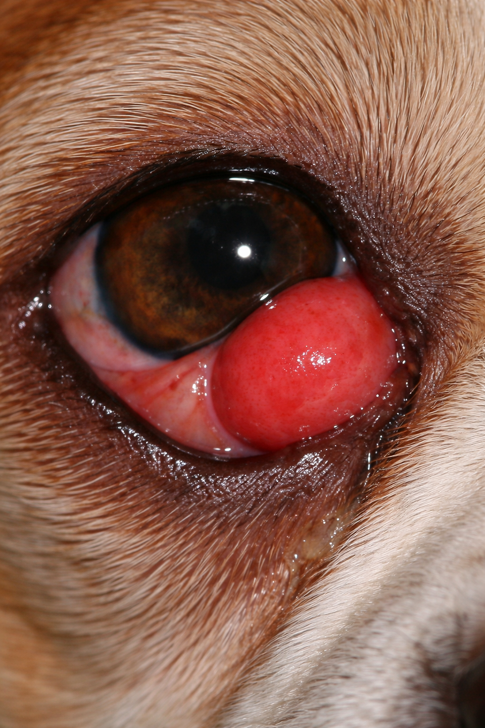
Cherry eye can affect one or both eyes and may appear suddenly or gradually. While the condition itself is not immediately life-threatening, it should never be considered harmless. The exposed gland becomes vulnerable to drying, inflammation, trauma, and secondary infections, all of which can compromise tear production over time.
If left untreated, cherry eye can lead to more serious complications such as:
Keratoconjunctivitis sicca (dry eye)
Chronic conjunctivitis
Corneal ulceration
Permanent damage to the tear-producing gland
From a clinical perspective, cherry eye is not simply a cosmetic issue. The long-term function of the gland is far more important than its appearance. For this reason, modern treatment approaches focus on preserving and repositioning the gland, rather than removing it.
It is also important to understand that cherry eye is primarily a structural and genetic condition, not an infectious disease. This means it cannot be prevented through hygiene alone and is more commonly seen in certain breeds with known connective tissue weaknesses.

Cost of Cherry Eye Treatment in Dogs (US & EU Price Breakdown)
The cost of treating cherry eye in dogs varies depending on several key factors, including location, clinic standards, surgical technique, and whether complications are present. Because cherry eye often requires surgical correction, understanding the full cost structure is essential for dog owners.
Average Cost Overview
What Affects the Cost?
Several variables can significantly influence the total expense:
Surgical technique used
Advanced methods (e.g., pocket technique) may cost more but have better outcomes
Clinic location and reputation
Urban and specialty clinics generally charge higher fees
Severity of the condition
Chronic or inflamed glands may require more complex procedures
Unilateral vs bilateral surgery
Treating both eyes increases total cost but may be more efficient in one session
Anesthesia and monitoring
Safer anesthesia protocols increase cost but reduce risk
Post-operative medications
Includes antibiotics, anti-inflammatory drugs, and artificial tears
Additional Hidden Costs
Owners should also consider indirect or follow-up expenses:
Recheck examinations
Protective collars (E-collar)
Repeat surgery in case of recurrence
Long-term tear supplements if gland function decreases
Cost vs Outcome Insight
While some owners may look for cheaper alternatives, it is important to understand:
Low-cost gland removal (obsolete method) may lead to permanent dry eye
Proper surgical repositioning preserves tear production and reduces long-term costs
In the long run, a well-performed surgery is more cost-effective than repeated treatments or complications.

Common Symptoms of Cherry Eye in Dogs
Cherry eye in dogs is usually easy to recognize, especially in its classic presentation. However, early or mild cases can sometimes be overlooked, making it important to understand the full range of clinical signs.
The most common symptom is:
A round, red or pink mass in the inner corner of the eye
This mass may vary in size and can appear intermittently at first before becoming permanent. In some dogs, the gland may prolapse only during stress or excitement and then temporarily retract.
Beyond this hallmark sign, several additional symptoms may be observed:
Visible Eye Changes
Swelling in the inner eyelid
Increased redness of surrounding tissues
Thickening of the third eyelid
Discharge (clear, mucoid, or purulent in secondary infections)
Behavioral Signs
Frequent eye rubbing or pawing
Squinting or partial eye closure
Sensitivity to light (photophobia)
Restlessness due to discomfort
Tear Film and Moisture Changes
Excessive tearing (epiphora) in early stages
Reduced tear production over time if gland function declines
Sticky or dry ocular surface in chronic cases
Secondary Complications
Conjunctivitis (inflamed conjunctiva)
Corneal irritation or ulceration
Increased risk of bacterial infections
In bilateral cases (both eyes affected), symptoms may appear asymmetrical, with one eye showing more severe prolapse than the other.
A key clinical point is that pain is not always prominent in early stages, which can mislead owners into delaying treatment. However, as the condition progresses, discomfort and complications become more likely.
Early recognition of these symptoms significantly improves treatment outcomes, especially when surgical correction is performed before chronic damage to the gland occurs.
Causes of Cherry Eye in Dogs
Cherry eye in dogs develops primarily due to structural weakness in the connective tissues that anchor the third eyelid gland in place. This weakness allows the gland to prolapse outward, becoming visible as the characteristic red mass.
Unlike infectious eye conditions, cherry eye is not caused by bacteria or viruses. Instead, it is a multifactorial condition, with genetics playing the most dominant role.
Genetic Predisposition
The most significant factor in cherry eye development is hereditary connective tissue weakness. Certain breeds are genetically predisposed to weaker anchoring ligaments around the third eyelid gland. In these dogs, even minor stress or normal eye movement can lead to gland prolapse.
This is why cherry eye is often seen:
At a young age (typically under 2 years)
Without any obvious trauma or trigger
Recurrently, even after temporary resolution
Weakness of the Orbital Ligament
The gland of the third eyelid is normally held in place by a fibrous ligament structure. When this ligament is:
Underdeveloped
Structurally weak
Degenerated over time
…the gland can easily slip out of its normal position.
This anatomical instability is the core mechanism behind cherry eye.
Inflammation and Secondary Irritation
Although not a primary cause, ocular inflammation can contribute to or worsen cherry eye. Conditions such as:
Conjunctivitis
Allergic eye reactions
Environmental irritants (dust, smoke)
…can lead to swelling in the eye tissues, increasing pressure and making gland prolapse more likely.
Trauma and Mechanical Factors
Direct or indirect trauma may trigger cherry eye in susceptible dogs:
Eye rubbing due to irritation
Rough play or minor injury
Sudden increases in intraocular pressure (straining, coughing)
However, trauma alone rarely causes cherry eye in dogs with strong connective tissue. It typically acts as a trigger in already predisposed individuals.
Age and Developmental Factors
Cherry eye is most commonly observed in:
Puppies and young dogs (under 1–2 years)
This is because their connective tissues are still developing and may lack full structural strength. Early onset is a strong indicator of genetic involvement.
Bilateral Risk
Dogs that develop cherry eye in one eye have a high probability of developing it in the other eye over time. This further supports the theory that the condition is systemic (genetic/anatomical), not localized.
Breeds Prone to Cherry Eye in Dogs
Certain dog breeds have a significantly higher risk of developing cherry eye due to inherited anatomical characteristics. These breeds often have looser connective tissues, shallow eye sockets, or prominent eyes, all of which contribute to gland instability.
High-Risk Breeds Table
Breed | Risk Level | Explanation |
Bulldog (English & French) | High | Weak connective tissue and characteristic facial structure |
Cocker Spaniel | Genetic predisposition affecting gland anchoring | |
High | Commonly reported in young individuals | |
Lhasa Apso | High | Shallow orbits and ligament weakness |
High | Brachycephalic anatomy increases risk | |
Pekingese | High | Prominent eyes and loose eyelid structure |
Boston Terrier | High | Compact skull and eye prominence |
Cane Corso | Moderate–High | Large breed with connective tissue susceptibility |
Neapolitan Mastiff | High | Heavy facial folds and weak support tissues |
Bloodhound | Moderate–High | Loose skin and eyelid laxity |
Basset Hound | Moderate–High | Droopy eyelids and connective tissue weakness |
Moderate | Occasional genetic predisposition | |
Moderate | Less common but still reported |
Key Observations
Brachycephalic breeds (short-nosed dogs) are at the highest risk
Dogs with loose skin and droopy eyelids are more susceptible
Large and giant breeds may also be affected due to connective tissue structure
Clinical Insight
From a practical standpoint, when a young dog from a high-risk breed presents with eye redness, cherry eye should be one of the first differential diagnoses.
Additionally, breeders and owners of predisposed breeds should be aware that:
The condition is often not preventable
Early intervention significantly improves outcomes
Surgical correction is commonly required in high-risk breeds
Types of Cherry Eye in Dogs (Partial vs Complete Prolapse)
Cherry eye does not always present in the same way. Understanding the different types helps determine the urgency of treatment and the most appropriate management approach.
Partial Prolapse
In partial prolapse:
The gland is not fully displaced
The red mass may appear intermittently
It can sometimes retract temporarily
Characteristics:
Mild swelling
Smaller visible mass
Symptoms may fluctuate
Often seen in early stages
Clinical Importance:
Partial prolapse is often underestimated. However:
It frequently progresses to full prolapse
Early intervention may improve surgical success rates
Complete Prolapse
In complete prolapse:
The gland is fully displaced and constantly visible
The mass is prominent and persistent
Characteristics:
Bright red, round swelling
Does not retract on its own
Often accompanied by irritation and discharge
Clinical Importance:
Higher risk of gland damage
Increased likelihood of secondary infections
Surgical treatment is almost always required
Unilateral vs Bilateral Cases
Cherry eye can also be classified based on the number of eyes affected:
Unilateral: Only one eye is affected
Bilateral: Both eyes are affected (may occur simultaneously or over time)
A key clinical observation:
Dogs with unilateral cherry eye have a high probability of developing it in the second eye later
Acute vs Chronic Cases
Type | Description | Clinical Impact |
Acute | Recently developed prolapse | Better surgical prognosis |
Chronic | Long-standing condition | Higher risk of gland damage and dry eye |
Clinical Insight
From a treatment perspective:
Early-stage (partial/acute) cases offer the best outcomes
Chronic or complete prolapse increases complication risk
Delayed treatment reduces the likelihood of full gland function recovery
Recognizing the type of cherry eye is critical for:
Choosing the correct treatment
Predicting prognosis
Preventing long-term ocular damage
Treatment Options for Cherry Eye in Dogs
Treatment of cherry eye focuses on restoring the gland to its normal position while preserving its function. Modern veterinary practice strongly emphasizes gland preservation rather than removal.
Surgical Treatment (Gold Standard)
Surgery is the most effective and commonly recommended treatment.
Pocket Technique (Most Preferred)
The gland is repositioned and secured within a conjunctival pocket
Preserves tear production
Low recurrence rate when performed correctly
Anchoring Technique
The gland is sutured to surrounding structures
Used in specific cases or when pocket technique is not suitable
Key Advantages of Surgery:
Restores normal anatomy
Prevents long-term complications
Maintains tear production
Why Gland Removal Is Not Recommended
In the past, the gland was sometimes removed. This approach is now considered outdated and risky.
Removal can lead to:
Chronic dry eye (KCS)
Lifelong need for eye medications
Increased risk of corneal damage
Preserving the gland is essential for long-term ocular health.
Medical (Non-Surgical) Management
Medical treatment alone does not cure cherry eye but may be used in specific situations:
Very early or mild cases
Temporary reduction of inflammation before surgery
Patients not suitable for anesthesia
Common Medical Approaches:
Anti-inflammatory eye drops
Lubricating artificial tears
Antibiotics (if infection is present)
However:
These treatments do not reposition the gland permanently
Relapse is almost inevitable without surgery
Manual Repositioning (Temporary)
In some cases, gentle manual pressure may temporarily reposition the gland.
Effect is usually short-lived
High recurrence rate
Not a definitive solution
Treatment Timing
Early intervention is critical:
Improves surgical success rates
Reduces risk of gland damage
Prevents chronic inflammation
Delaying treatment can lead to:
Fibrosis of the gland
Reduced tear production
Increased surgical difficulty
Clinical Decision Summary
Treatment Option | Effectiveness | Long-Term Outcome |
Surgery (Pocket) | Very High | Best outcome |
Surgery (Anchoring) | High | Good outcome |
Medical Management | Low | Temporary relief only |
Gland Removal | Not recommended | High risk of complications |
Step-by-Step Surgical Procedure for Cherry Eye in Dogs
Surgical correction of cherry eye is the gold standard treatment, aimed at repositioning and preserving the third eyelid gland. Among the available techniques, the pocket method is the most widely used due to its high success rate and low complication risk.
Below is a simplified, clinically accurate overview of how the procedure is performed:
Preoperative Preparation
Before surgery:
The dog undergoes a general health check
Tear production may be measured (Schirmer test)
The eye is examined for ulcers or infections
Fasting is required prior to anesthesia
This stage ensures the patient is safe for anesthesia and reduces surgical risks.
Anesthesia
The procedure is performed under general anesthesia
Local anesthetic drops may also be applied
The dog is positioned to allow optimal access to the eye
Safe anesthesia protocols are critical, especially in brachycephalic breeds.
Surgical Steps (Pocket Technique)
The third eyelid is gently everted (turned outward)
Two parallel incisions are made on the conjunctival surface
A “pocket” is created between the tissue layers
The prolapsed gland is carefully repositioned into this pocket
The incisions are closed with fine absorbable sutures
This technique hides the gland internally while preserving its function.
Duration of Surgery
Typically 15–30 minutes per eye
Bilateral cases may be completed in a single session
Immediate Postoperative Care
After surgery:
The dog is monitored until fully awake
Eye drops (antibiotic + anti-inflammatory) are prescribed
An Elizabethan collar (E-collar) is required
The collar is essential to prevent rubbing or trauma to the surgical site.
Success Rate and Recurrence
Success rate: 85–95% (depending on technique and case)
Recurrence risk: Low but possible, especially in severe or chronic cases
If recurrence occurs, a second surgery may be required.
Clinical Insight
The goal of surgery is not cosmetic correction, but functional preservation of tear production. Proper technique and early intervention significantly improve long-term outcomes.
Non-Surgical Management of Cherry Eye in Dogs
While surgery is the definitive treatment, non-surgical approaches may be used in specific situations. However, it is crucial to understand that these methods do not provide a permanent solution.
When Non-Surgical Management Is Considered
Very early-stage (mild, intermittent prolapse)
Temporary management before surgery
Patients unfit for anesthesia
Owner preference (with informed consent)
Medical Treatment Options
Anti-inflammatory Eye Drops
Reduce swelling of the gland
May temporarily decrease the size of the prolapse
Artificial Tears (Lubricants)
Maintain eye moisture
Protect the cornea from dryness
Antibiotic Drops
Used if secondary infection is present
Manual Repositioning
Gentle pressure may temporarily push the gland back into place
Often performed by a veterinarian
However:
Effect is usually short-lived
Recurrence is very common
Limitations of Non-Surgical Treatment
Method | Effect | Duration |
Eye drops | Reduces inflammation | Temporary |
Lubricants | Protects surface | Supportive only |
Manual reposition | Repositions gland | Very short-term |
Risks of Delaying Surgery
Relying only on medical management can lead to:
Chronic inflammation
Gland enlargement and fibrosis
Reduced tear production
Increased risk of dry eye (KCS)
Risks and Complications of Cherry Eye in Dogs
Although cherry eye surgery is generally safe and effective, like any medical procedure, it carries certain risks. Additionally, untreated or poorly managed cherry eye can lead to serious long-term complications.
Surgical Risks
Even with proper technique, the following complications may occur:
Recurrence of prolapse
The gland may prolapse again, especially in severe or chronic cases
Suture irritation
Internal sutures may cause mild irritation or inflammation
Infection
Postoperative infections are rare but possible
Swelling and inflammation
Temporary swelling is common in the first few days after surgery
Overcorrection or displacement
In rare cases, improper positioning can affect eyelid function
Long-Term Complications (If Untreated)
Leaving cherry eye untreated poses a much greater risk than surgery:
Keratoconjunctivitis sicca (Dry Eye)
Due to reduced tear production from gland damage
Chronic conjunctivitis
Persistent inflammation of the eye
Corneal ulcers
Resulting from dryness and irritation
Permanent gland damage
Loss of function due to prolonged exposure
Risk Factors for Complications
Certain factors increase the likelihood of complications:
Delayed treatment
Chronic or long-standing prolapse
Breed predisposition
Poor postoperative care
Inadequate surgical technique
Recurrence Rate Insight
Factor | Recurrence Risk |
Early surgery | Low |
Chronic cases | Moderate |
Poor technique | High |
High-risk breeds | Moderate–High |
Clinical Insight
The biggest mistake is underestimating cherry eye as a cosmetic issue. The real risk lies in loss of tear production, which can permanently affect eye health.
Early, proper surgical intervention significantly reduces all major risks.
Recovery Process After Cherry Eye Surgery in Dogs
The recovery period after cherry eye surgery is usually straightforward, but proper care is essential for a successful outcome.
Immediate Postoperative Period (First 24–48 Hours)
Mild swelling and redness are normal
The dog may show slight discomfort
Eye discharge may be present
At this stage:
Medications should be started as prescribed
The dog must wear an E-collar at all times
First Week After Surgery
Swelling gradually decreases
The gland remains in place if healing is successful
Sutures begin to stabilize the tissue
Owner responsibilities:
Administer eye drops regularly
Prevent rubbing or scratching
Monitor for abnormal signs (excess discharge, severe redness)
2–3 Weeks Post-Surgery
Most healing is complete
Sutures (if absorbable) begin to dissolve
Eye appearance returns closer to normal
At this stage:
Follow-up examination is recommended
E-collar may be removed if approved
Full Recovery Timeline
Stage | Timeframe | What to Expect |
Initial healing | 1–3 days | Swelling and mild discomfort |
Stabilization | 7–10 days | Reduced inflammation |
Functional recovery | 2–3 weeks | Normal gland position |
Full recovery | 3–4 weeks | Complete healing |
Signs of Successful Recovery
No visible prolapse
Normal tear production
Clear, moist eye surface
No signs of pain or irritation
Warning Signs (Require Attention)
Reappearance of red mass
Persistent swelling after 1 week
Yellow/green discharge
Excessive squinting or pain
Long-Term Outcome
With proper surgical technique and care:
Prognosis is excellent
Most dogs recover fully without complications
Tear production is preserved
When to See a Vet for Cherry Eye in Dogs
Timely veterinary intervention is essential for preventing permanent damage.
Immediate Veterinary Attention Required
Sudden appearance of a red mass in the eye
Persistent swelling or irritation
Eye discharge (especially yellow or green)
Signs of pain (squinting, pawing, sensitivity to light)
Post-Surgery Warning Signs
After treatment, seek veterinary care if:
The prolapse returns
Swelling worsens instead of improving
The dog cannot keep the eye open
There is excessive tearing or dryness
Routine Monitoring
Even if symptoms appear mild:
Early-stage cherry eye can worsen quickly
Monitoring both eyes is important
Follow-up exams improve long-term outcomes
Clinical Insight
Delaying veterinary care is one of the most common causes of complications. Early intervention offers:
Higher surgical success rates
Lower recurrence risk
Better preservation of tear function
Long-Term Prognosis of Cherry Eye in Dogs
The long-term outlook for dogs with cherry eye is generally very good, especially when treated early and appropriately.
Prognosis with Surgery
High success rate (85–95%)
Normal tear production preserved
Minimal long-term complications
Most dogs return to normal life without any lasting issues.
Prognosis Without Treatment
If left untreated:
Increased risk of dry eye (KCS)
Chronic inflammation
Corneal damage
Reduced quality of life
Factors Affecting Prognosis
Factor | Impact on Outcome |
Early treatment | Excellent prognosis |
Chronic cases | Reduced success rate |
Surgical technique | Critical for success |
Post-op care | Strong influence on recovery |
Breed predisposition | Moderate impact |
Bilateral Cases
Dogs with one affected eye often develop cherry eye in the other
Early monitoring allows faster intervention
Clinical Insight
Cherry eye is one of the few conditions where timing directly affects long-term function. Early surgical correction offers the best chance for a full recovery.
FAQ
What is cherry eye in dogs and why does it happen?
Cherry eye in dogs is the prolapse of the third eyelid gland, which normally sits hidden inside the lower inner corner of the eye. This gland is responsible for producing a significant portion of the tear film. The condition occurs when the connective tissue that holds the gland in place becomes weak, allowing it to protrude outward. This is most commonly due to genetic predisposition rather than infection or trauma. Certain breeds are more prone, and it often appears at a young age without any obvious trigger.
Is cherry eye in dogs painful?
Cherry eye is not always immediately painful, especially in early stages. However, it does cause discomfort and irritation. Dogs may rub their eyes, squint, or show sensitivity to light. As the condition progresses, the exposed gland becomes more vulnerable to drying and inflammation, which can lead to pain, infections, or even corneal damage. So while it may start as a mild issue, it can become painful if left untreated.
Can cherry eye in dogs go away on its own?
In most cases, cherry eye does not resolve permanently on its own. Sometimes the gland may temporarily move back into place, especially in early or mild cases, but recurrence is very common. Without proper treatment, the condition usually persists or worsens over time. Medical treatments like eye drops may reduce swelling temporarily, but they do not fix the underlying structural problem.
Do all dogs with cherry eye need surgery?
Most dogs with cherry eye will eventually require surgery, especially if the prolapse is persistent. Surgery is considered the most effective and long-term solution because it repositions the gland and preserves its function. Non-surgical treatments may be used in early or mild cases, but they rarely provide a permanent fix. Delaying surgery can increase the risk of complications such as dry eye.
Is cherry eye surgery safe for dogs?
Yes, cherry eye surgery is generally safe and widely performed. When done by an experienced veterinarian, the success rate is high, typically between 85–95%. As with any procedure involving anesthesia, there are some risks, but these are usually minimal. Proper preoperative assessment and postoperative care significantly reduce complications.
How long does cherry eye surgery take and what is the recovery time?
The surgery itself usually takes about 15–30 minutes per eye. Recovery typically takes 2–3 weeks, with most dogs returning to normal within a month. During this period, eye drops are used, and an E-collar is required to prevent the dog from rubbing the eye. Follow-up checks are important to ensure proper healing.
Can cherry eye come back after surgery?
Yes, recurrence is possible but relatively uncommon when proper techniques are used. The risk of recurrence depends on factors such as the surgical method, the severity of the condition, and the dog’s breed. If recurrence occurs, a second surgery may be needed. Early intervention and correct technique significantly reduce this risk.
What happens if cherry eye is not treated?
If left untreated, cherry eye can lead to serious complications. The most important risk is reduced tear production, which can result in dry eye (keratoconjunctivitis sicca). This condition can cause chronic irritation, infections, and even vision problems. Over time, the gland may become permanently damaged, making treatment more difficult.
Is cherry eye contagious to other dogs?
No, cherry eye is not contagious. It is not caused by bacteria or viruses but by anatomical and genetic factors. Therefore, it cannot spread from one dog to another.
Can cherry eye affect both eyes?
Yes, cherry eye can affect both eyes. In many cases, dogs that develop cherry eye in one eye may eventually develop it in the other. This is especially common in genetically predisposed breeds. Monitoring both eyes is important even if only one is currently affected.
Which dog breeds are most at risk for cherry eye?
Breeds such as Bulldogs, Cocker Spaniels, Beagles, Shih Tzus, Lhasa Apsos, and other brachycephalic or loose-skinned breeds are at higher risk. These breeds often have structural characteristics that make the gland more prone to prolapse.
Can cherry eye be prevented?
Cherry eye cannot be fully prevented because it is largely genetic. However, early detection and prompt treatment can prevent complications. Avoiding eye irritation and maintaining good overall eye hygiene may help reduce triggers but will not eliminate the risk entirely.
Is cherry eye surgery expensive and is it worth it?
The cost varies depending on location and clinic, but it is generally considered a worthwhile investment. Surgery not only corrects the visible problem but also preserves tear production, preventing more serious and costly conditions in the future. In most cases, early surgery is more cost-effective than managing long-term complications.
Will my dog’s vision be affected by cherry eye?
Cherry eye itself does not directly affect vision. However, if left untreated and complications develop—such as corneal ulcers or dry eye—vision can be affected over time. This is why early treatment is important.
What should I do if I notice a red mass in my dog’s eye?
You should seek veterinary attention as soon as possible. Early diagnosis and treatment improve outcomes and reduce the risk of complications. Avoid trying to treat or manipulate the eye at home, as this can worsen the condition.
Keywords
cherry eye in dogs, dog eye prolapse, third eyelid gland dog, cherry eye treatment dog, cherry eye surgery cost dog
Sources
Source | Link |
American College of Veterinary Ophthalmologists (ACVO) | |
Merck Veterinary Manual | |
American Veterinary Medical Association (AVMA) | |
VCA Animal Hospitals | |
Mersin Vetlife Veterinary Clinic |